Science as art
A photo exhibition shows the beauty born in Princeton’s labs and field research
PRINCETON’S ART OF SCIENCE exhibition, a showcase of 44 images representing science as an art form, will be on display at the Liberty Science Center in Jersey City, N.J, through Sept. 15. The exhibit draws from five years of photographs in the University’s Art of Science competition, with images selected by renowned photographer and professor emeritus Emmet Gowin and former Princeton University Art Museum curator Joel Smith *01. The photos spotlight the striking aesthetic and scientific importance of Princeton’s research, from fluid mechanics to biological fieldwork to plasma physics. “It’s special to have your research highlighted as art,” says Steve Brunton *12, a featured photo winner from 2010, “and it’s a different way to express yourself than writing peer-reviewed papers.” The competition was sponsored by the David A. Gardner ’69 Fund in the Council of the Humanities and the School of Engineering and Applied Science.
Vivienne Chen ’14, an English major, is a freelance writer and videographer who contributes frequently to PAW and PAW Online.
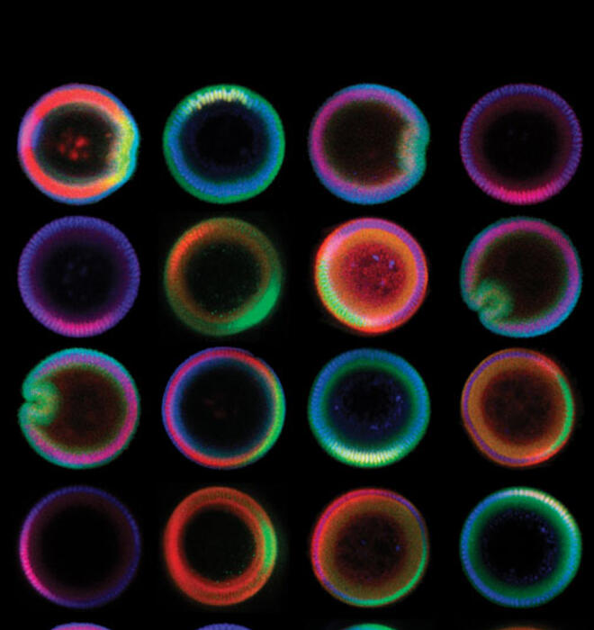
Patterning the Embryo (above)
Yoosik Kim *11, Stanislav Shvartsman
For Yoosik Kim *11 and Professor Stas Shvartsman, a chance detour to an art exhibit inspired the presentation of their scientific research. “We were studying the cross sections of the fruit-fly embryo,” says Shvartsman, a professor of chemical and biological engineering who supervised Kim, “but after looking at a large number of these embryos, we found out there was a Kandinsky exhibition at the Guggenheim.” Russian abstract painter Wassily Kandinsky’s iconic paintings of colorful concentric circles, known as Kandinsky circles, captivated the researchers. “Once I saw these paintings,” says Shvartsman, “I knew we needed to arrange our embryos in the same way.” Their photo shows how the embryo is subdivided into three main tissue types — muscle, skin, and nerve — creating a rainbow of cross sections arranged like Kandinsky circles.
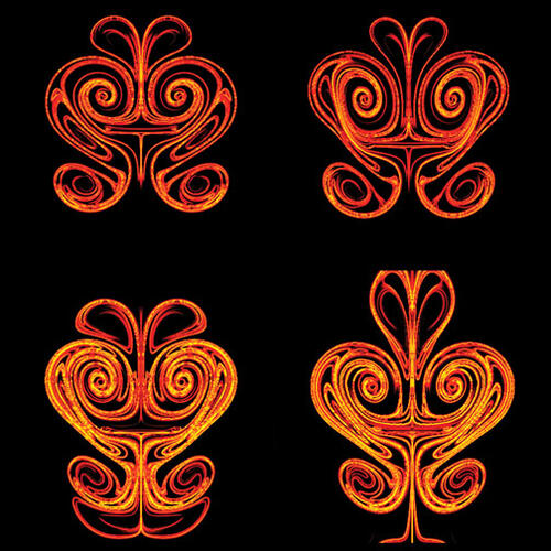
Stirring Faces
Steve Brunton *12
Steve Brunton *12 was testing some new computational tools during his graduate studies on fluid flow when he created these computer-generated swirls. “What’s exciting about this research is that even with a simple input, we get complex structures in the flow field,” says Brunton, “and it’s amazing to watch that development in real time.” The patterns that emerged when simulating a rigid wing in a still fluid — like a fruit fly flapping its wings, Brunton explains — were striking to him for their symmetry and complexity. Brunton even made large prints of his photo for his son’s bedroom.
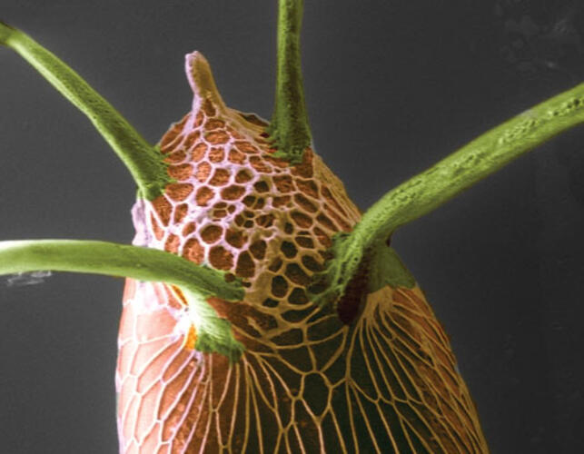
Five-Horned Eggshell
Nir Yakoby, Maria Pia Rossi
The “horns” in Nir Yakoby and Maria Pia Rossi’s photo are not horns at all: The four green stalks actually are tubes extending from a fruit-fly egg, which Yakoby explains are used like “snorkels” to help the egg breathe after it is laid in a fruit. The white horn is used for the male fly to deposit sperm. “The egg itself is about 300 microns in size,” says Yakoby, a Rutgers University assistant professor who was a postdoc in Princeton’s Lewis-Sigler Institute for Integrative Genomics when this photo was taken. “It’s a speck if you look at it with your eye.” But with a scanning electron microscope and some post-production artificial coloring, the egg becomes the giant, intricate organ in the photograph, which Yakoby and Pia Rossi, an applications engineer, examined during their research. “We want to learn how cells decide what to become,” Yakoby says.
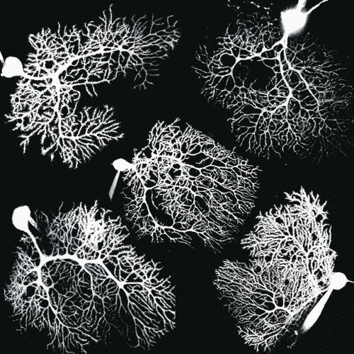
Purkinje Neurons
Dmitry Sarkisov *06
This collection of snapshots of the branch-like structure in the brain cells of rats came from Dmitry Sarkisov *06’s research on long-term plasticity in the cerebellum. The white neurons are lit by a calcium-sensitive dye, which Sarkisov used to examine the cells micrometer by micrometer during his thesis work. The beauty of the photo, Sarkisov says, is in how the “geometry represents their unique function.” The cells “receive inputs from tens of thousands of neurons,” he explains, thus requiring the web of dendrites leading to the circular mass of the cell body.
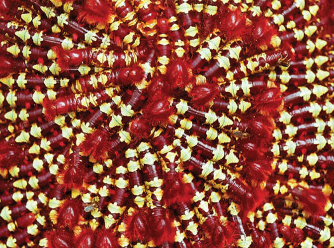
No Sibling Rivalry Here
Trond H. Larsen *06
Trond Larsen *06’s photo of a swarming caterpillar colony was captured during his Ph.D. research in the Lago Guri islands of Venezuela. “I remember it was a particularly hot day, over 100 degrees,” Larsen says. “I noticed these caterpillars on the underside of a leaf above my head while I was cutting a new trail into an unexplored area of one of the islands. ... I was struck by the bright, distinctive coloration of each individual caterpillar, but even more so by the colors and design that emerges from the entire group.” Larsen, now on the staff of the group Conservation International, explains that the tight clustering of the caterpillars creates a confusing image that may deter predators — either by disguising their identity or by implying toxicity with their bright patterns. “Scientifically, it is fascinating to try to understand how this group behavior evolved,” he says.






No responses yet