Breaking ground - Chemical engineering
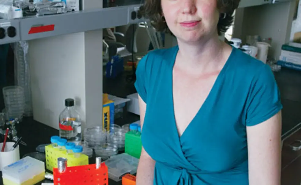
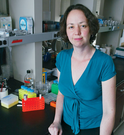
Nelson, who earned her Ph.D. in biomedical engineering from Johns Hopkins, aims to combine engineering and biology to enhance the basic understanding of how tissues grow and to discover how they react when development goes awry.
Her primary interest is in branched tissues, the treelike structures in lungs, kidneys, and mammary glands. “We try to understand how that tree forms — how millions and millions of cells know to build themselves into such a complex architecture,” Nelson says.
Nelson and her colleagues build tissues in culture using microfabrication techniques similar to those used in the semiconductor industry. But instead of placing inorganic components on a flat computer chip, they organize living cells in a three-dimensional tissue structure. Nelson also uses computer models to simulate the development of organic tissues.
In one project, Nelson’s lab is working on a model mammary gland that simulates tissue fibrosis, a process in which cells produce an excess of a polymer called extracellular matrix that builds up and increases the stiffness of the tissue. The tissue change can make women more susceptible to breast cancer, and a reliable model of fibrosis eventually could be used to screen for therapies to combat the process.
To create that model, Nelson must answer several basic questions about how fibrotic cells behave: How do cells respond to alterations in the stiffness of their surroundings? What signals cause cells to begin fibrosis? The model, when complete, could help to answer other elusive questions, like whether or not normal cells can overwhelm the detrimental effects of a single fibrotic cell.
There are few standard techniques for building and studying a developing tissue, according to Nelson. Embryolo-gists have centuries of knowledge about whole organs, and biologists have sophisticated techniques for manipulating single cells. But the “in-between” area has not been explored thoroughly.


No responses yet